Global and local similarity of the primary visual cortex: mechanisms of orientation preferenceDavid M. Alexander1, Phil Sheridan2, Paul D. Bourke1, Otto Konstandatos3
The authors wish to thank Jim Wright, Clare Chapman, Geoff Stuart,
1
Brain Dynamics Laboratory,
Mental Health Research Institute, Melbourne,
Vic, Australia
AbstractThe local-global similarity model of V1 proposes two transformations to derive the organisation of orientation preference. Both these transforms are mappings of the hemi-retinal image, and are achieved by known connectivity in the visual system. The transformations are a global retinotopic mapping into the supragranular layers and a tiling of the supragranular layers with multiple local versions of the hemi-retinal image. The local mapping implies that the lateral connections in lamina 4B preserve the topographic ordering of the visual hemi-field. Since the local mapping of the supragranular layers produces a simultaneous expansion and contraction of the current visual inputs, it accounts for both the local response properties for orientation preference and their global geometric organisation. The interaction of the two topographically identical mappings is shown to predict the formation of patchy intrinsic connections in the supragranular layers. The mappings described allow those properties of the visual field which can be predicted on the basis of spatial contiguity (texture, orientation, colour, contrast) to be available as response properties over the entire visual field.
1 IntroductionThis paper presents a model of the mammalian primary visual cortex. The model assumes that the geometry of V1 serves to organise multiple response properties into two cortical dimensions. The core proposition is the existence of two, topographically identical mappings of the visual field to the primary visual cortex, which define the geometrical organisation of orientation preference and drive the formation of patchy connectivity in the supragranular layers.
The local-global similarity (LGS) model proposes that V1 is doubly-similar in the following sense. Input from the eye reaches the supragranular layers of V1 via two mappings: a global mapping and a local mapping. Multiple copies of the local mapping tile the area of V1. The global mapping supplies retinotopic information to the upper layers. Each local mapping (via interactions with the global mapping and other local maps) supplies additional response properties such as orientation preference. The LGS model of the primary visual cortex suggests there is a direct relationship between globally represented objects (e.g. an oriented line) and various other response properties (e.g. orientation preference) which have a local geometry.
The function of the LGS mapping is to provide each point in the retinotopic image with a representation of the entire visual hemi-field. Each retinotopic point is able to learn a `snapshot' of points in the visual image whose activity tends to coincide with that retinotopic point. In the case of orientation preference, the set of oriented lines passing through a retinotopic point can be associated with that point.
2 Response Property Geometry of V1The response properties of V1 are well catalogued. These properties have been mapped through single-cell studies (Hubel and Wiesel, 1968), metabolic transport studies (Tootell et al, 1988a; 1988b; 1988c) and optical imaging of the cortical surface (Blasdel, 1992; Blasdel and Salama, 1986). These studies have shown that V1 exhibits a distinctive geometrical tiling (Blasdel, 1992). Swindale (1996) has described a set of canonical properties which a model of the geometry of the primary visual cortex should take into account. These properties include the spatial frequencies and organisation of: ocular dominance bands, cytochrome oxidase (CO) blobs, singularities and iso-orientation regions. To Swindale's list may be added: spatial frequency selectivity, patchy intrinsic connectivity and cortical point spread. Layer 4C of the macaque primary visual cortex has a strict retinotopic organisation (Blasdel and Fitzpatrick, 1984). This layer receives the vast bulk of inputs from the retina, via the thalamus (Blasdel and Lund, 1983). In the macaque, each hemi-retina is distorted, through a complex logarithmic mapping, to an almond shaped mapping in layer 4C of V1 (Tootell et al, 1988a). This distortion is of fundamental importance to the functionality of the visual recognition system, allowing computational simplification for operations such as rotation and scaling in two dimensions (Schwartz, 1980; Sheridan and Alexander, 1997). Receptive field studies (Hubel and Wiesel, 1977) reveal an orderly, though less exact retinotopic organisation in the supragranular layers. Receptive field sizes are also larger in the supragranular layers. Neurons in the upper layers of the primary visual cortex are organised into repeated units, roughly 800 um wide and 600 um high, called hypercolumns (Hubel and Wiesel, 1968). Each hypercolumn spans an entire range of orientation tunings and a left and right ocular dominance set. Located along the centres of ocular dominance bands are cytochrome-oxidase (CO) blobs. The blobs appear to CO staining because of their higher metabolic activity (Horton and Hubel, 1980), and are responsive to colour, high contrast and low spatial frequencies. Interblob regions are more selective for low contrast and high spatial frequencies (Tootell et al, 1988b; Tootell et al, 1988c). The geometry of orientation preference reveals three predominant features: singularities, linear zones and saddle regions (Blasdel, 1992). Orientation preference changes continuously around points, or singularities. These roughly circular `pinwheels' traverse 180o of all possible orientation preference. Between adjacent singularities, running parallel to the ocular dominance bands, are regions in which orientation preference changes slowly and continuously. These regions are called linear zones. Other regions between singularities show local minima of orientation preference in orthogonal directions: so-called saddle points. Singularities surrounding saddle-points form mirror images of each other, through both a vertical reflection line and a horizontal one. The double reflections have the effect of allowing orientation selectivity to change continuously across the edges singularities. Singularities and CO blobs both tend to lie along the centres of ocular dominance bands (Bartfield and Grinvald, 1992; Livingstone and Hubel, 1984). In most cases CO blobs lie in between singularities, although in up to 15% of cases singularities and CO blobs coincide (Bartfield and Grinvald, 1992). It is not known whether the incidence of CO blob and singularity overlap varies over the surface of V1, for example, whether it is more prevalent in the foveal region.
3 Connectivity of Granular and Supragranular Layers
Anatomically, the mapping into layer 4C occurs via the Lateral Geniculate
Nucleus (LGN) in the thalamus
(Blasdel and Lund, 1983). In layer 4C, two
streams of inputs from the magnocellular and parvocellular layers of the LGN
are recombined to form an orderly retinotopic representation
(Blasdel and Fitzpatrick, 1984).
The inputs from the LGN project to layer 4C in discrete
blocks. Projections from the parvocellular layers of the LGN form dense
terminal boutons in lamina 4C
The supragranular layers receive little or no input directly from 4C, but
receive the bulk of their inputs via lamina 4A and 4B
(Blasdel et al, 1985).
Lamina 4C Of particular interest for the model presented in this paper are the lateral projections of lamina 4B spiny stellates within 4B itself. These efferent axons project laterally up to 4.5 mm in the macaque (Blasdel et al, 1985). A similar pattern of projecting fibres is found in the squirrel monkey (Rockland and Lund, 1983). The lateral connections show periodic accumulations of denser terminal fibres every 375-400 um (Blasdel et al, 1985; Rockland and Lund, 1983). These patches of connections form a radial pattern with a similar spatial periodicity to the CO blobs (Rockland and Lund, 1983). The fibres within 4B extend further than any other class of intrinsic fibres within the primary visual cortex. Only cells within layer 4B send out this class of long range lateral fibres and, at mid- to long-range distances from the cell body, all terminating boutons are within 4B itself. Most of the fibres within 4B are preferentially horizontal, rather than vertical (Rockland and Lund, 1983). The set of connections within 4B is ideally suited to carry out the mapping, proposed in this paper, of the retinotopic representation in the granular layers to a local mapping in the supragranular layers. The chief requirement of this mapping is that each coarse-grained (~ 400 x 300 um) retinotopic location in lamina 4C connects to a point in every local map (~ 400 x 300 um) in the supragranular layers. This mapping occurs through polysynaptic pathways within lamina 4B. Studies using injections of retrograde and antereograde tracer have revealed underlying regularities in patchy intrinsic connections within the supragranular layers of V1 (Blasdel et al, 1985; Bosking et al, 1997; Malach et al, 1993; Rockland and Lund, 1983; Hubel and Wiesel). These connections traverse the grey matter parallel to the cortical surface in the supragranular layers and project to discrete patches or targets, sometimes several millimetres from the site of tracer injection. Use of these tracer techniques in conjunction with other imaging techniques has revealed that patchy connections tend to prefer targets with the same response properties. This has been shown for orientation selectivity and ocular dominance (Malach et al, 1993) and CO/interblob zones (Yoshioka et al, 1996). The spatial pattern of these patchy intrinsic connections is therefore closely related to the spatial pattern of other response-property systems discussed thus far.
Recent work on the postnatal development of area 17 in the Ferret has revealed that the patchy intrinsic connections form from an initially diffuse set of random connections in the upper layers (Ruthazer and Stryker, 1996). The formation of patchy connections occurs before the presence of visual input and before the appearance of response properties associated with the supragranular layers, for example orientation selectivity. We therefore assume these connections reflect some primary transformation of early visual inputs, upon which later fine tuning of response properties is based. This paper describes two mappings of visual inputs through the granular and supragranular layers, and how the interaction of the two mappings drives the formation of patchy intrinsic connections in the supragranular layers.
It has recently been shown, in the primary visual cortex of the tree shrew,
that the patchy intrinsic connections are not perfectly radial, but form an
elongated pattern
(Bosking et al, 1997). The axis of elongation corresponds to
the preferred orientation of the injection site. In other words, if tracer is
injected into a site in the supragranular layers with a preferred orientation
of
4 Local-Global Similar mappingAlexander et al (1997) have noted strong analogies between the global properties of the hemi-retinal image and the response properties in the supragranular layers. These are given in table 1. In particular, we assume these analogies with the hemi-retinal image apply to a geometrical unit in the supragranular layers corresponding to 1/4 of a hypercolumn. Such a unit includes one CO blob and one singularity and has the approximate dimensions in the macaque of 400 um x 300 um (Blasdel, 1992).
The present model of V1 introduces a few simplifications into the visual system, and will not explicitly deal with ocular dominance, the cortical magnification factor, spatial frequency, contrast and colour selectivity. The resulting simplified model focuses on the development of orientation selectivity, and aims to explain data from mammals such as the macaque, the tree shrew and the ferret. A more complete model is in development.
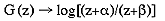
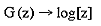
The global retinotopic input-mapping from the hemi-retinal image to the granular layers (in particular layer 4C) can be approximated by the function of a complex variable supplied by Schwartz (1980):
where
and
or
where
The retinotopic representation of lamina 4C most directly reaches the
supragranular layers via lamina 4C
Receptive field mapping has shown that the retinotopic receptive field
structure of the supragranular layers is not as exact at that in lamina 4C.
This fits with anatomical data suggesting somewhat diffuse inputs into the
supragranular layers via 4A and larger receptive fields for the upper layers
(Blasdel and Fitzpatrick, 1984).
For the sake of simplification, in this
description of the LGS model of V1, we assume that the retinotopic
input-mapping between lamina 4C
A second stream of inputs reaches the supragranular layers via laminae
4C
where
This mapping may also be expressed as
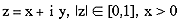
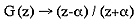
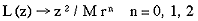
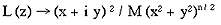
where M is a scaling factor which shrinks the size of the map (M >> 1). This mapping has the effect of mapping a semi-circle to a full circle. In the case of n=0, it maps equally spaced lines of iso-eccentricity to radii that increase as a square of the eccentricity. If n=1, equally spaced lines of iso-eccentricity are preserved. If n=2, the pattern described for n=0 is reversed. The mapping described in equation 5 is illustrated in diagram 2. A set of rays--of angles spanning 180o --converging on the fovea, is mapped by this function to a circular pinwheel. Multiple copies of this input-mapping tile the supragranular layers. According to Schwartz (1980) there are approximately 3,000 hypercolumns in the macaque primary visual cortex. This leads to an estimate of 12,000 tilings of the local mapping described by equation 5. The most critical feature of the mapping is that it is a local mapping. It allows each retinotopic point to be associated with a local representation of the whole visual field. The next most important feature of this mapping is that it maps a semi-circle to a circle, doubling the angles. This approximates, in the local mappings, the shape of experimentally imaged singularities. Anatomically, we propose the local input-mapping described by equations 5-7 occurs through the lateral connections formed by spiny stellates in lamina 4B (Rockland and Lund, 1983). These lateral connections recombine the retinotopic image in a series of steps to form the desired local input-mapping to the supragranular layers. This is consistent with the observations that 1) the longest lateral fibres are in 4B, and 2) most fibres within 4B terminate at great distances within 4B. This anatomy is consistent with the required one to all mapping. The relevant anatomy is summarised in diagram 3. The critical prediction of the LGS model is that the lateral connections in lamina 4B preserve the topology of the retinal image, while replicating that topology many-fold over the surface of V1. Each point on the supragranular layers can be defined in terms of a doubly-similar coordinate geometry. Each point is a function of the pair (zG,zL) where zG specifies, in discrete complex coordinates, which local input-map the point falls within, where
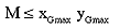
If there are M tilings of the local input-map, then
since the rounded shape of the primary visual cortex means it does not fill the complex plane of zG entirely. The term zL specifies the coordinates of the point within that local input-map. Since this point is a target neuron, zL is also in discrete complex coordinates. The definition of the point (zG,zL) in the supragranular layers is illustrated in diagram 4. We express the inverse of the global input-mapping function (e.g. the inverse of equation 1) as G-1(z) and the inverse of the local input-mapping function (e.g. the inverse of equation 5) as L-1(z). Each point, (zG,zL), in the supragranular layer receives input from two points in the visual field, one through the global retinotopic input-mapping, point g:
and another through the local input-mapping, point l:
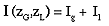
where g and l are complex variables and are points on the hemi-retina. The input into a given point of the supragranular layers, I (zG,zL), is simply
where Ig is the total afferent input from point g and Il is the total afferent input from point l.
5 Interaction Between MapsThe global input-mapping described by equations 1-4 supplies the retinotopic receptive field properties of the supragranular layers. Other response properties of the local map representations are formed through an interaction of the local input-maps with the global retinotopic input-mapping into the supragranular layers. Here we distinguish between the local input-map, supplied by equations 5-7 and the local representational maps (or local response property maps) which form as a result of interactions between the input maps, other cortical anatomy and hebbian learning driven by visual stimuli. The response properties of local representational maps form through hebbian learning mechanisms as a result of correlated neuronal activity. The LGS mapping allows each coarse-grained (~ 400 x 300 um) retinotopic input into the supragranular layers to become associated with a local mapping (also ~ 400 x 300 um) of the entire hemi-field. Since these maps are hypothesised to form only when both sets of inputs (i.e. Ig and Il in equation 12) are active, and even adjacent local maps receive different Ig's, each local representational map in the supragranular layers learns a different set of patterns. In the general case, each local map in the supragranular layer `sees' a different set of stimuli. For each Ig, the supragranular layer also receives inputs, Il, from every point in the visual hemi-field. Responses to these local mappings of the visual field are reinforced by activity from the retinotopic inputs Ig. Only those points in the visual field whose activity reliably coincides with the point g will form strong connections within the map driven by Ig. The local representation which forms will be a version of the visual field which is visually relevant to the Ig supplying retinotopic input to that local map. In the case of line orientation preference, each local map `sees' only the subset of line orientations relevant to that local map. The subset of line orientations which pass through the hemi-retinal point, g, comprises all the oriented lines which produce supragranular activity in the local map driven by that Ig. The local map, insofar as its response properties are due to the interaction of Ig and Il, will therefore learn responses to only the subset of oriented lines passing through the point g. Each local map learns a subtly different set of lines orientations, depending on its retinotopic location. The conjunction of the local mapping of an oriented line, and the global mapping of the point it passes through, is termed a point/orientation conjunction. A point/orientation conjunction describes the relevant pattern of activity, or representation, for line orientation preference. A critical feature of the input-mapping given in equations 5-7 is that it simultaneously performs both a contraction of the layer 4C representation to multiple local images in the supragranular layers and an expansion of the layer 4C representation so that any point in the hemi-field becomes spread out in the upper layer representation. That is, each local input, Il, reaches every version of the local map. This property leads to an explanation for the formation of patchy intrinsic connections in the supragranular layers. We assume that the transform described in equations 5-7 occurs from at least from the time of early post-natal development, that is, prior to earliest visual experience. We further assume that patchy connections in the upper layers are formed from an initial random connectivity. These assumptions are consistent with recent findings on the development of lateral connections in the ferret primary visual cortex (Ruthazer and Stryker, 1996). The lateral connections in the supragranular layers are refined through the detection of correlated activity, between adjacent local maps, in neurons receiving inputs from both local and global input-maps. This is illustrated in diagram 5. A short line is represented in both the global mapping of the supragranular layers and the local mappings which tile the supragranular layers. Where activity in the two adjacent local-map representations coincides, patchy connections form. Connections are assumed to form between any two of these representations which have correlated activity over time through a simple hebbian learning mechanism. It should be noted that patchy connections form between cortical points in the supragranular layers (i.e. between Il's) whose representations are spatially contiguous in layer 4C. The patchy connectivity therefore reflects the expansion of the cortical image implied by the multiple tilings of the map described by equations 5-7. Patchy connectivity maintains direct connectivity, within the local supragranular representation, between representations drawn from the adjacent (or the same) retinotopic locations in layer 4C. We propose that the mapping in equations 5-7 is critical to the development of lateral connections and also critical to their maintenance over time. However, once the response properties are acquired during this development process, moment to moment visual processing in V1 depends primarily on the interactions between the retinotopic representation in layer 4C and the supra-granular response properties supplied by the patchy intrinsic connections. If this is the case then visual processing V1 in is not critically slowed by the proposed poly-synaptic junctions in the transformation of the representation from layer 4C, through lamina 4B, into the supragranular layers. The mappings described in this paper reveal how the primary visual cortex recognises oriented lines in different retinotopic locations. The LGS mapping uniquely identifies any line by position and angle, since for a given orientation, there is only one line which passes though that retinotopic position. The patchy intrinsic connections link together the set of point/orientation conjunctions, each of which individually defines the line. The patchy connectivity therefore allows a further avenue of disambiguation, possibly adding a binding label through phase synchronisation of the neuronal activity (Singer, 1993). Synchronised activity between linked point/orientation conjunctions enhances response properties to oriented lines, thus generalising the response property to regions beyond a particular retinotopic location. In the general case, the model predicts that V1 can learn stimulus features which are predictable on the basis of spatial contiguity. The interaction, in the supragranular layers, between a specific retinotopic input and the local map inputs from the entire visual hemi-field leads to a unique pattern of activation at that retinotopic point. Properties such as line orientation, spatial frequency, contrast and colour tend to be spatial contiguous and so will reliably activate particular patterns of activity in adjacent local representations. Through this reliable co-activation, hebbian learning drives the formation of lateral connectivity between the active sites in these adjacent maps, producing generalised response properties. A few additional assumptions are required to reproduce the geometric pattern of orientation preference, at the scale of individual singularities, hypercolumns and beyond. To retrieve the typical singularity shape, of 180o of orientation preference traversing 360o of a circle, responses to oriented lines of a local map must favour orientations within a range of approximately 180o. This is the distortion provided by equation 5. Systematic mapping of direction preference in the primary visual cortex of the ferret reveals that direction preference is most often segregated into local maps with only a 180o range of direction preference (Weliky et al, 1996). Imaging of orientation preference shows that adjacent singularities are often mirror images of each other; this produces the saddle regions between sets of singularities. The tiling of multiple singularities shown in diagram 6 illustrates this mirroring, revealing multiple saddle points. Reflection of the adjacent tiles has the effect of allowing orientation preference to change continuously between singularities. It does not alter the functionality of the LGS mapping, which only relies on a local representation of the visual hemi-field being mapped to each global retinotopic point. Diagram 7 illustrates the relationships between CO blobs and singularities in the model. The model predicts that singularities and CO blobs should coincide in the region on the global retinotopic map which represents the centre of the field of vision. This is because the CO blobs are hypothesised to be the local map representations of the central field of vision and local-mappings in this region are activated by the set of orientations passing through the central field of vision. This contrasts to the local-map representations in the periphery. The set of orientations passing through these retinotopic locations miss the central field of vision and hence, in their local map representations, the CO blobs. This may explain the variation in findings in the literature, regarding the positioning of CO blobs and singularities (Bartfeld and Grinvald, 1992; Livingstone and Hubel, 1984).
A simplification in this analysis of orientation preference has been the nature
of the retinotopic inputs into the supragranular layer-hypothesized to be
connections from 4C
6 Conclusions
This paper introduces three assumptions to explain orientation selectivity in
the primary visual cortex. These are: a global, topology-preserving mapping
from the retina to the supragranular layers via the laminae
4C Three additional assumptions allow the model to predict the specific geometrical patterns of response properties for orientation preference. These assumptions are: that the local input-maps distort the hemi-retinal field so that polar angles are doubled; orientation responses are limited to a range of 180o within a particular local map; and, the tiling of the supragranular layers requires two reflections of the local input map. These assumptions allow the model to reproduce many aspects of the distinct geometry of orientation preference in the primary visual cortex. The LGS mapping of visual inputs allows those properties of the visual field which can be predicted on the basis of spatial contiguity (orientation, texture, colour, contrast) to be available as response properties over the entire visual field. The local mapping to the supragranular layers makes the contents of the entire visual field available for association with each retinotopic location in the supragranular layer. Patchy intrinsic connections link these response properties at different retinotopic locations. Further description of the model will treat direction preference, as well as simulations of the formation of patchy intrinsic connections (Alexander et al, ms. in prep.). Extension of the model to response properties such as colour selectivity and spatial frequency preference follow from the general case in a straight forward manner. The LGS mapping described by equations 5-7 bears correspondences to the ice-cube model of Hubel and Wiesel (1977). Both assume the uppers layers are tiled with a regular map of the feature space. The LGS mapping has the additional features that it 1) directly suggests the function of known anatomical connections, 2) explains how the response properties are created. The model assumes that the geometry of V1 serves to organise multiple response properties into two dimensions. It differs from other models of V1 geometry which, rather than mappings, use simulated annealing or other relaxation procedures to achieve this dimension reduction (Durbin and Mitchison, 1990; Swindale, 1992). The latter models have multiple free parameters (Swindale, 1996). By contrast, the LGS model of V1 contains a limited number of assumptions and no arbitrary parameters.
Alexander D.M., Sheridan P., Bourke P.D. and Wright J.J. (ms. in prep.) The local and global similarity of the primary visual cortex: simulation of direction preference. Alexander D.M., Sheridan P., Bourke P.D. (1997) An algebraic-geometric model of the receptive field properties of the macaque striate cortex, Proceedings of the Australian Neuroscience Society, 8, 62. Bartfeld E. and Grinvald A. (1992) Relationships between orientation-preference pinwheels, cytochrome oxidase blobs, and ocular-dominance columns in primate striate cortex, Proceedings of the National Academy of Sciences USA, 89, 11905-11909. Blasdel G.G. and Lund J.S. (1983) Termination of afferent axons in macaque striate cortex, Journal of Neuroscience, 3(7), 1389-1413. Blasdel G.G. and Fitzpatrick (1984) Physiological organization of layer 4 in macaque striate cortex, Journal of Neuroscience, 4(3), 880-895. Blasdel G.G., Lund J.S. and Fitzpatrick D. (1985) Intrinsic connections of macaque striate cortex: axonal projections of cells outside lamina 4c, Journal of Neuroscience, 5(12), 3350-3369. Blasdel G.G. and Salama G. (1986) Voltage-sensitive dyes reveal a modular organization in monkey striate cortex, Nature, 321, 579-585. Blasdel G.G. (1992) Orientation selectivity, preference, and continuity in monkey striate cortex, Journal of Neuroscience, 12(8), 3139-3161. Bosking W.H., Zhang Y., Schofield B. and Fitzpatrick D. (1997) Orientation selectivity and the arrangement of horizontal connections in tree shrew striate cortex, Journal of Neuroscience, 17(6), 2212-2221. Durbin R. and Mitchison G. (1990) A dimension reduction framework for understanding cortical maps, Nature, 343, 644-647. Horton J.C. and Hubel D.H. (1980) Regular patchy distribution of cytochrome oxidase staining in primary visual cortex of the macaque, Nature, 292, 762-764. Hubel D.H. and Wiesel T.N. (1968) Receptive fields and functional architecture of the monkey striate cortex, Journal of Physiology (London), 195, 215-243. Hubel D.H. and Wiesel T.N. (1977) Functional architecture of macaque monkey visual cortex, Proceedings of the Royal Society (B), 198, 1-59. Livingstone M.S. and Hubel T.N. (1984) Anatomy and physiology of a color system in the primate visual cortex, Journal of Neuroscience, 4, 309-380. Malach R., Amir Y., Harel M. and Grinvald A. (1993) Relationship between intrinsic connections and functional architecture revealed by optical imaging and in vivo targeted biocytin injections in primate striate cortex, Proceedings of the National Academy of Sciences USA, 90, 10469-10473. Miller R. (1996) Neural assemblies and laminar interactions in the cerebral cortex, Biological Cybernetics, 75, 253. Rockland K.S. and Lund J.S. (1983) Intrinsic laminar lattice connections in primate visual cortex, Journal of Comparative Neurology, 216, 303-318. Ruthazer E.S. and Stryker M.P (1996) The role of activity in the development of long-range horizontal connections in area 17 of the ferret, Journal of Neuroscience, 16(22), 7253-7269. Schwartz E.L. (1980) Computational anatomy and functional architecture of striate cortex: a spatial mapping approach to perceptual coding, Vision Research, 20, 645-669. Schwartz E.L. (1983) Cortical mapping and perceptual invariance: a reply to Cavanagh, Vision Research, 23 (8), 831-835. Sheridan P. and Alexander D.M. (1997) Invariant transformations on a space-variant hexagonal grid, Proceedings of Vision, Recognition, Action: Boston. Singer, W. (1993) Synchronization of cortical activity and its putative role in information processing and learning, Annual Review of Physiology, 55, 349-374. Swindale N.V. (1992) A model for the coordinated development of columnar systems in primate striate cortex, Biological Cybernetics, 66, 217-230. Swindale N.V. (1996) The development of topography in the visual cortex: a review of models, Network, 7, 161-247. Tootell B.H., Switkes E., Silverman M.S. and Hamilton S.L. (1988a) Functional anatomy of the macaque striate cortex. II. Retinotopic organization, Journal of Neuroscience, 8(5), 1531-1568. Tootell B.H., Silverman M.S., Hamilton S.L., Switkes E. and De Valois R. (1988b) Functional anatomy of the macaque striate cortex. V. Spatial Frequency, Journal of Neuroscience, 8(5), 1610-1624. Tootell B.H., Silverman M.S., Hamilton S.L., De Valois R. and Switkes E. (1988c) Functional anatomy of the macaque striate cortex. II. Color, Journal of Neuroscience, 8(5), 1569-1593. Weliky M., Bosking W.H. and Fitzpatrick D. (1996) A systematic map of direction preference in primary visual cortex, Nature, 379, 725-728. Yoshioka T., Levit J.B. and Lund J.S. (1994) Independence and merger of thalamocortical channels within macaque monkey primary visual cortex: anatomy of interlaminar projections, Visual Neuroscience, 11, 467-489. Yoshioka T., Blasdel G.G., Levit J.B. and Lund J.S. (1996) Relation between patterns of intrinsic lateral connectivity, ocular dominance, and cytochrome oxidase-reactive regions in macaque monkey striate cortex, Cerebral Cortex, 6, 297-310.
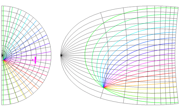
Global mapping of the hemi-retinal image into the supragranular layer of the primary visual cortex. This mapping is given in equation 1. Left of figure shows the hemi-retinal field which is mapped to the supragranular layers, shown in the right of figure. The distortion of the hemi-retinal image is shown by black lines. The point f, where the converging black lines meet, is the fovea and has the z coordinates (0,0). The mapping is global, that is, it covers in a single mapping the entire area of V1. The mapping of a set of oriented lines is shown by coloured lines. Each coloured line represents the set of moving lines, of a given orientation and of variable speed, which converge on the point g. In this representation, the distance of a moving bar from the point g is proportion to its speed.
Diagram 2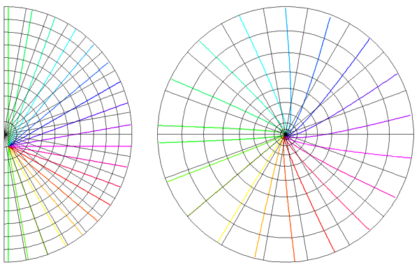
Local mapping of the hemi-retinal image into the supragranular layers of the primary visual cortex. This mapping is given in equation 1, n=0. Left of figure shows the hemi-retinal field which is mapped to the supragranular layers, shown at right of figure. The distortion of the hemi-retinal image is shown by black lines. The point f, where the converging black lines meet, is the fovea and has the z coordinates (0,0). The mapping is local, that is, it tiles the area of V1 with multiple small copies of the z-plane. The mapping of a set of oriented lines is shown by coloured lines. Each coloured line represents the set of moving lines, of a given orientation and of variable speed, which converge on the point l. In this representation, the distance of a moving bar from the point l is proportion to its speed.
Diagram 3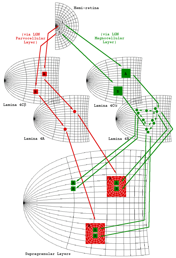
Anatomy of V1 relevant to the LGS model. Two
streams of inputs reach the supragranular layers. One mapping, via laminae
4C
Diagram 4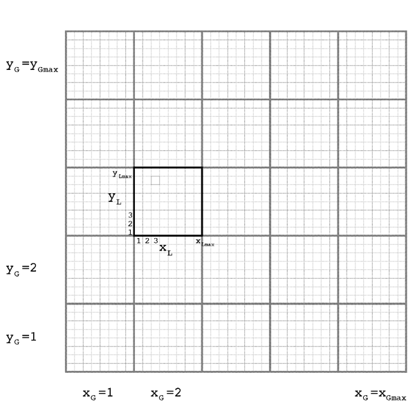
LGS coordinate geometry of the primary visual cortex. The coordinate system consists of a global complex plane, zG, which is tiled with multiple local complex planes, zL. The small black square has the coordinates xG = 2, yG = 3, xL = 3, yL = 7.
Diagram 5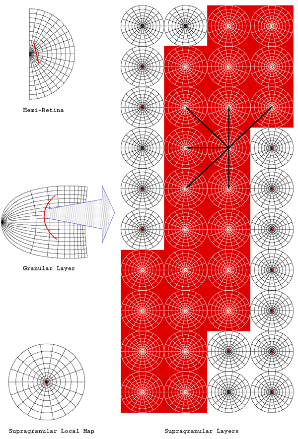
Schematic representation of the formation of intrinsic patchy connectivity. A short, oriented line (shown in red) is mapped according to the LGS model of V1. The left hand column shows the mapping from hemi-retina to the granular and supragranular layers. Right of diagram shows a magnified section of the line and the intersecting local maps. Each point/orientation conjunction uniquely defines the line by position and angle. Patchy connectivity forms between pairs of the point/orientation conjunctions, through a hebbian learning rule (shown by black arrows).
Diagram 6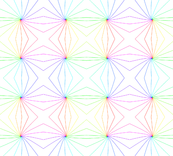
Tiling of the supragranular layers with multiple copies of the hemi-retinal image. Adjacent tilings are reflected about either a vertically or horizontally oriented axis. The tiling shows saddle points between singularities.
Diagram 7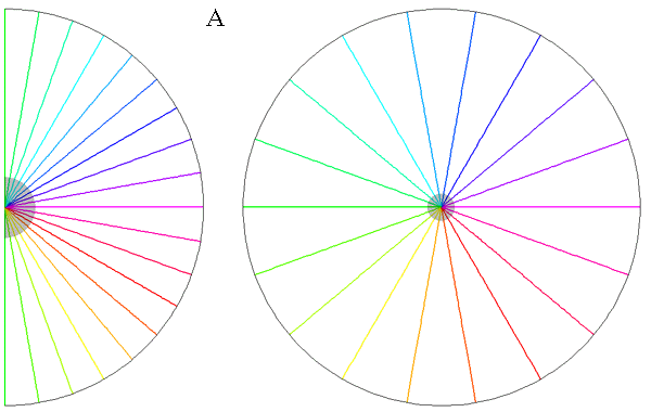 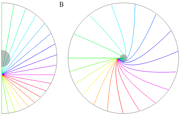
Relationships between singularities (colour) and
CO blobs (grey).
|
 .
The size of these dense terminating patches is
approximately the width of an ocular dominance column, 400 um. The terminating
input patches to 4C
.
The size of these dense terminating patches is
approximately the width of an ocular dominance column, 400 um. The terminating
input patches to 4C in finer patches of approximately 100 um.
 ,
the pattern of patchy intrinsic connections overlies a
global, retinotopic representation of a line passing through that point and
having an orientation
,
the pattern of patchy intrinsic connections overlies a
global, retinotopic representation of a line passing through that point and
having an orientation